Ideas and discoveries
Doctors and technicians focus on radiation exposure after a nuclear disaster or the effectiveness of radiotherapy

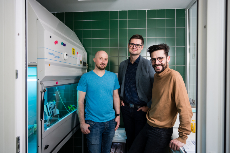
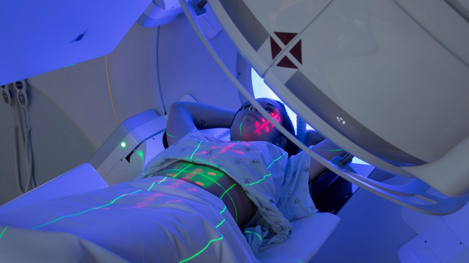
A head or neck cancer diagnosis is bad in itself, unfortunately even radiation treatment is not a certain cure, as it does not work for a large proportion of patients. Subsequent surgery after failure of radiotherapy becomes much more complicated for surgeons, as wounds in a previously irradiated person heal poorly. But what if it were possible to predict the effectiveness of radiotherapy before it started? The software that assists doctors and scientists in this endeavour was developed at the BUT in cooperation with the CAS and MU.
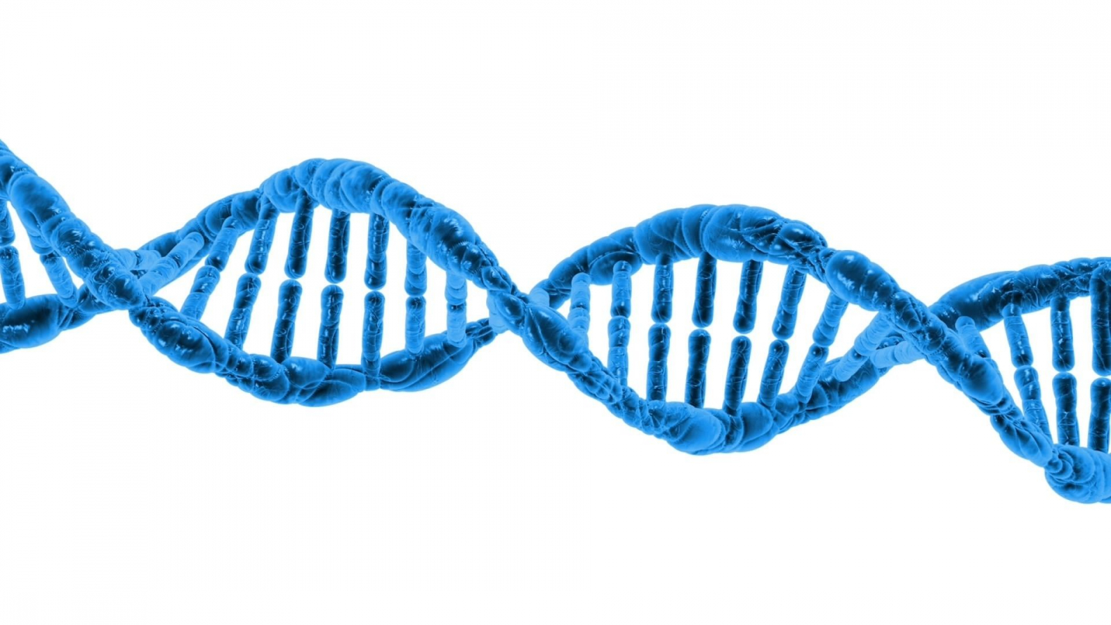
“DNA has two strands and when both break, the molecule breaks, which is one of the most serious types of damage that can be inflicted on this vital molecule. Ionising radiation is very effective in creating these double-strand DNA breaks, which is why it is used in radiotherapy, where it can effectively kill cancer cells. We can mark and image these DNA breaks under a microscope,” explains Martin Falk, the head of the Department of Cell Biology and Radiobiology at the Institute of Biophysics of the Czech Academy of Sciences. He also adds that cells have a variety of mechanisms and proteins involved that, when cells are irradiated, “rush” to the fracture areas to try to repair the damage as quickly as possible. Scientists can image these proteins using a fluorescent dye.

Coloured foci (speckles) of glowing proteins marking DNA breaks that can be observed under a microscope are the main protagonists of the research conducted at the BUT by Tomáš Vičar from the Department of Biomedical Engineering, Faculty of Electrical Engineering and Communication. His task was to create a programme that can automatically find and count these places, determining as many shape and other parameters as possible. “We tried standard methods, but traditional algorithms failed due to the high variability of the studied foci both over time and between different cell types or even individual samples. In addition, we wanted to work with 3D cell images, which is much more complicated than the situation in 2D. Artificial intelligence methods are currently very popular for image processing. Instead of telling the program that it should count a focus if the intensity of the displayed DNA break is greater than a certain value, our algorithm learns this on its own,” Vičar emphasises the importance of artificial neural networks.
Although the team had to feed the software with training data for a year and a half, it is now functional and fully autonomous. Besides the two proteins studied so far, it even learned to analyse foci of other proteins on its own. Martin Falk of the CAS admits that humans are still more reliable in some respects: “When a person looks at the same images of damaged cells for 15 years, they are probably still more accurate than a piece of software. But when large amounts of data need to be analysed, manual analysis is virtually impossible, and especially in practice, the speed of obtaining results, which is enormous compared to a human, is important to us.”
And how much time will the program save for doctors? “We examine one sample manually for several days to a week; the machine can do it in about an hour. Moreover, it does not take people’s time and the calculation can run at night,” says Tomáš Vičar about one of the solution’s biggest advantages.

The team from the CAS, Masaryk University and BUT examine both purposeful irradiation in cancer treatment and harmful effects on human health during accidental irradiation. This could happen to astronauts, for example, who are exposed to completely different types of radiation in space than on Earth. However, scientists also work with catastrophic scenarios, such as a nuclear power plant accident. In such a case, blood would be drawn on the spot from persons affected by radiation, processed and microscopic images of the blood cells analysed by computer. They would quickly determine out who needs priority care because they were more strongly affected by the radiation.
However, a more likely use is assessing the effectiveness of radiotherapy in cancer treatment. The software could examine both pre- and post-treatment patient samples. If the programme shows that the blood sample does not respond to radiation, doctors would not start radiotherapy and would not burden the patient with a treatment that would be useless in that patient’s case. On the contrary, they might start a more effective treatment strategy right away.
Experts have the opportunity to work with samples of real patients thanks to the inclusion of several institutions, which is ensured by Jaromír Gumulec from the Masaryk University, Faculty of Medicine: “We have been monitoring patients for about 6 years and therefore know how they responded to radiotherapy. Our question then is simple – does what we predicted using our software correspond to how patients responded to the actual treatment?”
(tk)
Scientists from FEEC BUT are participating in the development of a smart intersection. This could make transport more efficient and ensure greater safety
YSpace succeeds in prestigious ESA programme and heads to space
Scientists from FEEC BUT have developed a bracelet that warns of the risk of Parkinson ’s disease
Fake or real signature? FEEC scientists develop software for handwriting forensics
Swarm of unmanned drones with ground robots to help army survey hazardous areas