Topic
Villain or Hero? The Electron Microscope as a Detective
While Brno may not rival the size of other European metropolises, if you place it on the map of global microscopy, you’ll see that it stands up to the magnetic pull of the proverbial Mecca. Brno is a powerhouse in electron microscopy, and CEITEC plays a significant role in this field. The Brno Electron Microscopy Days provide CEITEC with an opportunity to open its doors to the public and participate in the program with lectures, guided tours, and demonstrations of cutting-edge technologies used at the centre to study materials and living cells down to the nanometer scale.
Brno: the city of microscopes and innovation
Over the past century, Brno has established itself as a hub for electron microscopy. Today, roughly one-third of the world's electron microscopes are produced in this region, and local scientists and engineers rank among the best in the field. Alongside industrial giants such as Thermo Fisher Scientific and TESCAN, Brno is also home to numerous scientific institutions, with CEITEC standing out as a leading research centre.
CEITEC as a centre of excellence
CEITEC provides a unique environment for cutting-edge research in electron microscopy. Its laboratories house state-of-the-art instruments that enable scientists to study structures with resolution down to individual atoms. Researchers here are also dedicated to methodological innovations that further push the boundaries of imaging technology.
One of CEITEC’s key research approaches combines cryo-electron microscopy with correlative techniques, allowing for the detailed study of biological samples while preserving their natural state. This method is crucial for investigating biomolecular complexes and their roles in diseases such as cancer and neurodegenerative disorders.
With the help of electron microscopes, CEITEC scientists tackle the mysteries of cellular stress, uncover how viruses infiltrate cells, and contribute to solving antibiotic resistance. These microscopes also reveal material structures at the atomic level, analyse and modify their chemical composition, and even engrave patterns using electron beam lithography—a technique essential for producing next-generation semiconductor chips.
Scientific collaborations and applications
CEITEC collaborates with industrial partners and academic institutions worldwide. Thanks to its top-tier infrastructure and expertise in imaging technologies, it is a crucial partner in materials research, nanotechnology, and biomedicine. In the field of new nanomaterials, revolutionary approaches are being developed for disease diagnostics and treatment. CEITEC is also actively engaged in semiconductor research and plays a key role in the establishment of the planned National Semiconductor and Chip Centre, set to launch this year.
Collaboration is also a driving force within the Brno region itself—local players in electron microscopy have joined forces to create a platform working to ensure that when people around the world hear "electron microscopy," the first place they think of is Brno. The city thrives on microscopy, especially in the spring, when banners and events for the Brno Electron Microscopy Days pop up all over town.
Electron Microscopy Days at CEITEC
During the Electron Microscopy Days (link only in Czech), the public has a unique opportunity to discover how microscopy shapes scientific progress. Leading electron microscope manufacturers and research institutions welcome visitors of all ages into their facilities, offering programs that highlight innovations and discoveries in the microscopic world (link only in Czech).
As every year, CEITEC will host guided tours of its laboratories, where experts will demonstrate the practical applications of electron microscopy in research. Visitors can listen to lectures on current trends in the field and discuss with researchers involved in the latest scientific discoveries, or expand their knowledge of the laws of physics or see how light interacts with matter.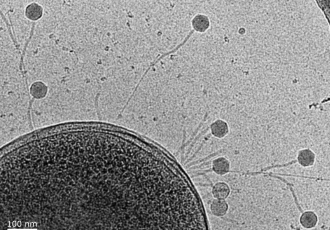
At CEITEC BUT (Brno University of Technology) in the technology park, visitors can explore how materials appear under a microscope—revealing fascinating structures such as collagen foams, cartilage replacements, and even nanoparticles. Meanwhile, at CEITEC MUNI (Masaryk University) in the Bohunice campus, they can meet the facility’s four-meter-tall giant—Titan Krios, an advanced electron microscope used to study the structures of viruses. Visitors will also be able to hold 3D models of viruses, offering a tangible insight into the microscopic world.
During the Electron Microscopy Days, both science centres will give people a glimpse into the world of microscopic "villains" and "heroes". And although electron microscope images are originally black and white, the microscopic world of heroes and villains is not so simple. The villains can be, for example, pathogens that cause disease and the heroes can be beneficial microorganisms or new biomaterials with therapeutic potential, but e.g. the physics experiments at CEITEC BUT will introduce you to electrons in both roles.
The future of electron microscopy at CEITEC
As the demand for highly detailed analysis of materials and living cells continues to grow, CEITEC is constantly evolving and expanding its capabilities. The future likely lies in greater automation and the integration of electron microscopy with artificial intelligence, paving the way for faster and more precise analyses. Brno will thus maintain its status as a global leader in electron microscopy, with CEITEC playing a key role in shaping its future.
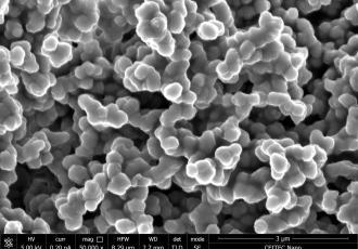
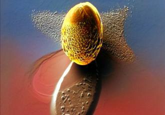
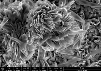
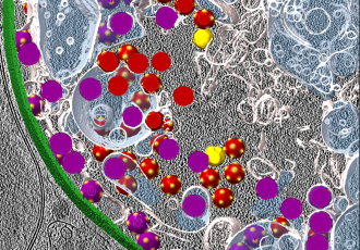
In the photo gallery, you’ll find images of materials and microorganisms captured by CEITEC scientists using electron microscopy.
Source: CEITEC, author: Halina Jílková

CEITEC Scientists Aim to Increase "High-Speed Microscopy" Up to Fivefold
Microscope of the Future: Combining Light and Electrons Wins Prestigious Grant
100 years since the birth of Josef Dadok, pioneer of instrumentation technology
In Brno she found her second home and her dream job. A theoretical scientist from Iran shares her perspective on life in the Czech Republic
A priceless offer. CEITEC will help researchers to bring the results of their work to the market