Ideas and discoveries
3D cartilage atlas of mid-trimester human embryos
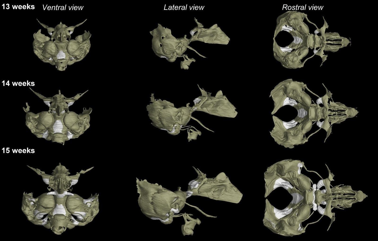
Our knowledge of human body development is often studied in animal model organisms. Scientists from the Laboratory of X-ray Micro- and Nano-Computed Tomography at CEITEC BUT have long been collaborating with biologists around the world and in previous studies have shown the development of, for example, cartilaginous tissue (chondrocranium) in mouse embryos, which is a crucial structure for the further development of the skull as well as other tissues in the developing organism. There are many ways to study animal organisms. In comparison, the possibilities for studying human embryos are very limited, both in terms of methodology and ethical issues. Current research, therefore, often has to rely on drawings made by anatomists a few decades ago.
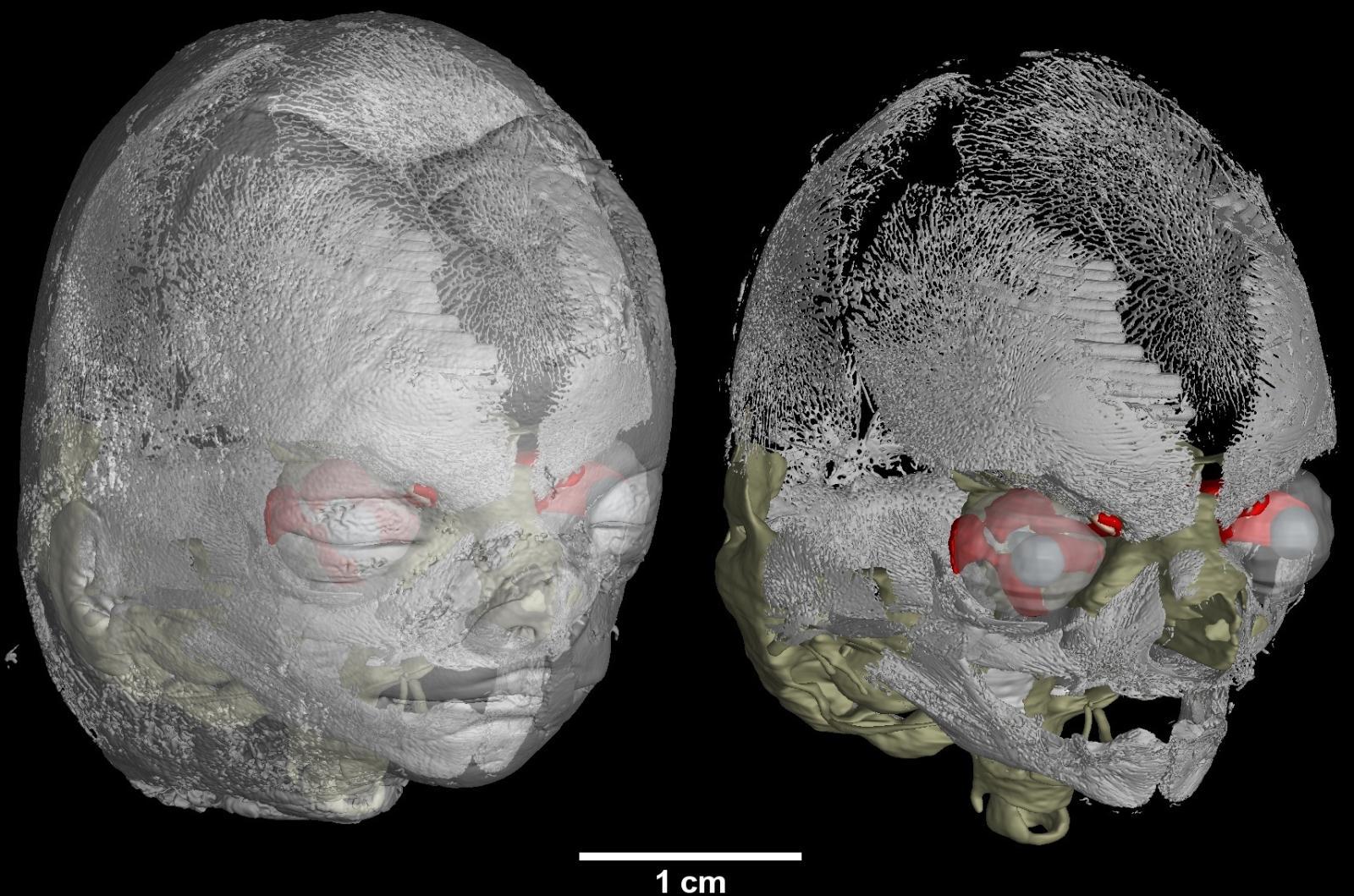
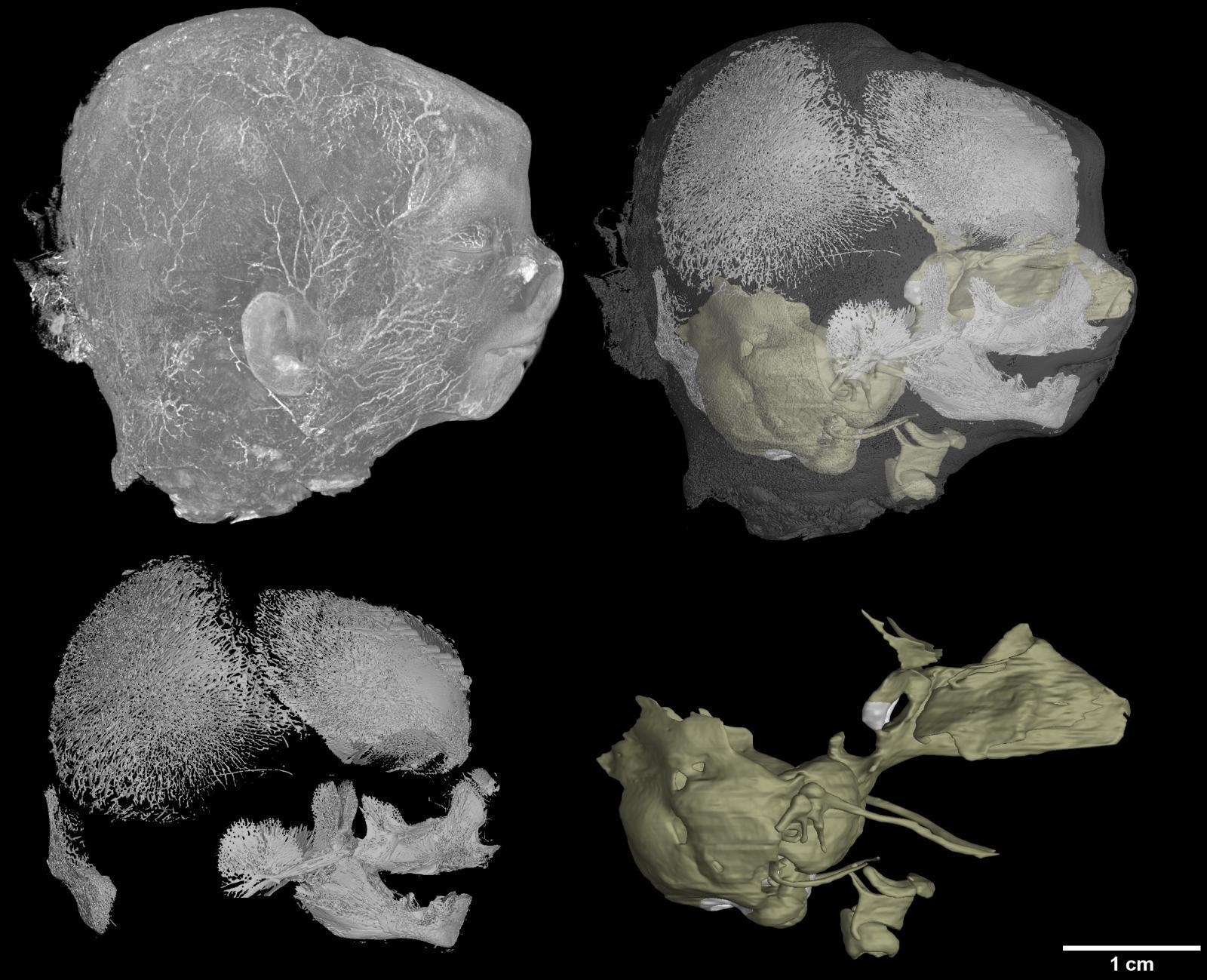
Scientists from the X-ray Micro- and Nano-Computed Tomography Laboratory at CEITEC BUT joined forces with an international team of biologists and the University of British Columbia (UBC) in Canada, which works closely with a local hospital and had human embryos in its collection. These samples were collected in the 1980s by Dr. Virginia Diewert, professor emerita at UBC. These were embryos of elective or spontaneous terminations, where the mother gave anonymous consent for the use of the foetus for scientific purposes. The embryos from this collection were taken to Brno, where they were first prepared so that soft tissues, such as cartilage, could be imaged by X-ray. This was followed by scanning by X-ray computed tomography and then demanding data processing, focusing not only on the cartilage but also on the ossified areas and other soft tissues.
There are several collections of human embryos in the world, mostly from the last century. However, hospitals and universities alike are aware of how precious the material is, and gaining their trust to measure such rare samples is very difficult. „In our case, our long-term cooperation with Marcela Buchtová from the Czech Academy of Sciences proved helpful. She spent some time at the UBC under the supervision of Professor Joy Richman. Professor Richam had tried to scan samples in Canada, but she had not achieved sufficient data quality to visualize the internal structure. When she saw our results with the mouse embryos, she decided to try to join forces. Our credibility was further strengthened by the fact that we had already published some papers with Prof. Igor Adameyko, who is among the leading figures in developmental biology and who was also involved in the research,“ said Markéta Kaiser, a Technician in the field of Physics at CEITEC BUT.
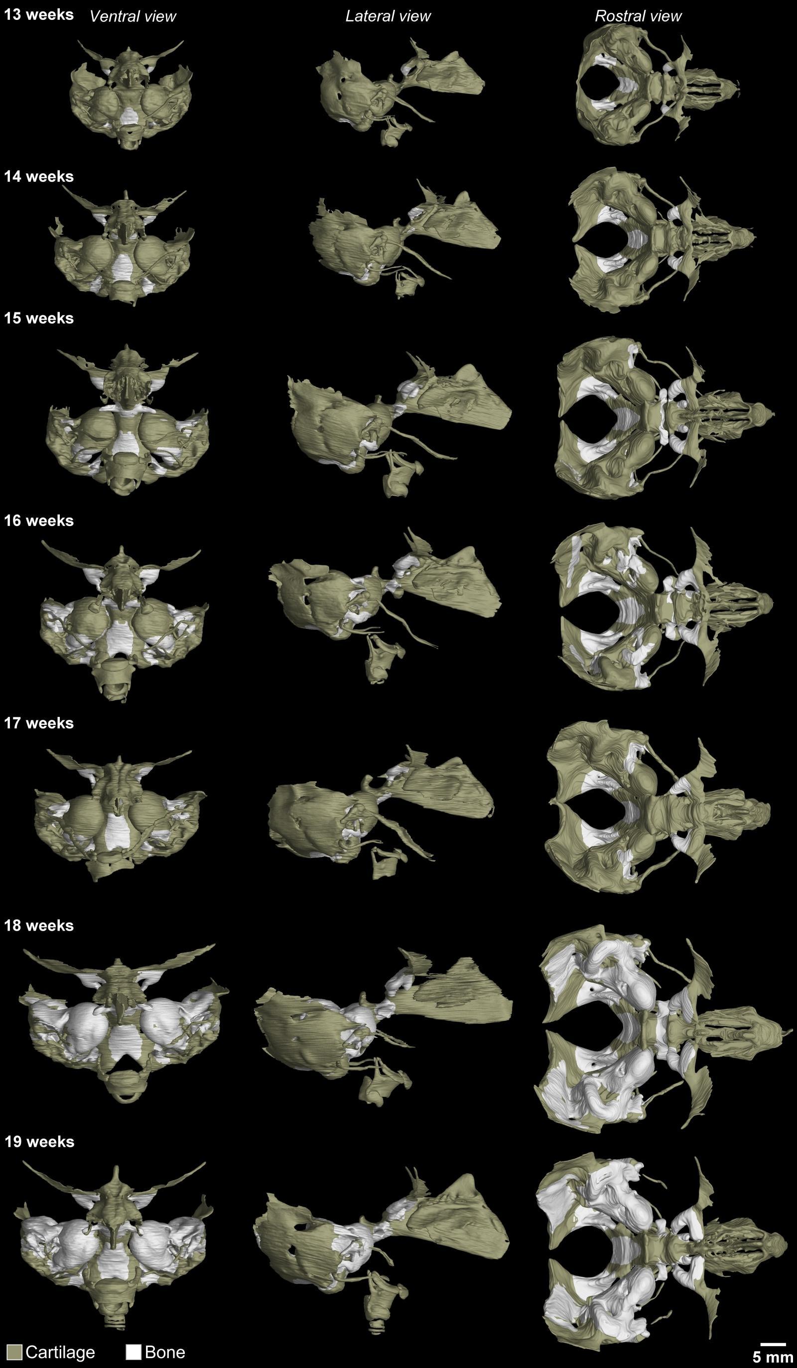
The new study "3D atlas of the human fetal chondrocranium in the middle trimester" published in Nature Scientific Data provides the first comprehensive 3D atlas of the developing cartilage of human embryos in the second trimester of pregnancy. The study of cartilaginous structures in the foetal skull is important for understanding normal and abnormal foetal development. The knowledge gained will be applicable to developmental biology, clinical research, and prenatal diagnosis. In the future, the resulting 3D atlas could help improve prenatal examination of foetal morphology and potential treatment strategies.
Using X-ray computed tomography is a non-destructive way to look inside a sample without damaging it. In our case, after the measurements were taken, all the embryos were sent back to Canada and placed in the hospital’s collection, where they can await their next opportunity to help us understand the development and function of the human body.
Source: CEITEC BUT
Brno is an oasis of peace for me, I feel relaxed here, says Chinese doctoral student Xia
Awarded student Victory Jaques has developed a method to preserve researched microsamples of cultural heritage
Science is made by people and people are driven by passion. However, passion alone is not enough, says Vinicius Santana
The memory of Josef Dadok lives on at CEITEC through cutting-edge research in spectroscopy
Villain or Hero? The Electron Microscope as a Detective